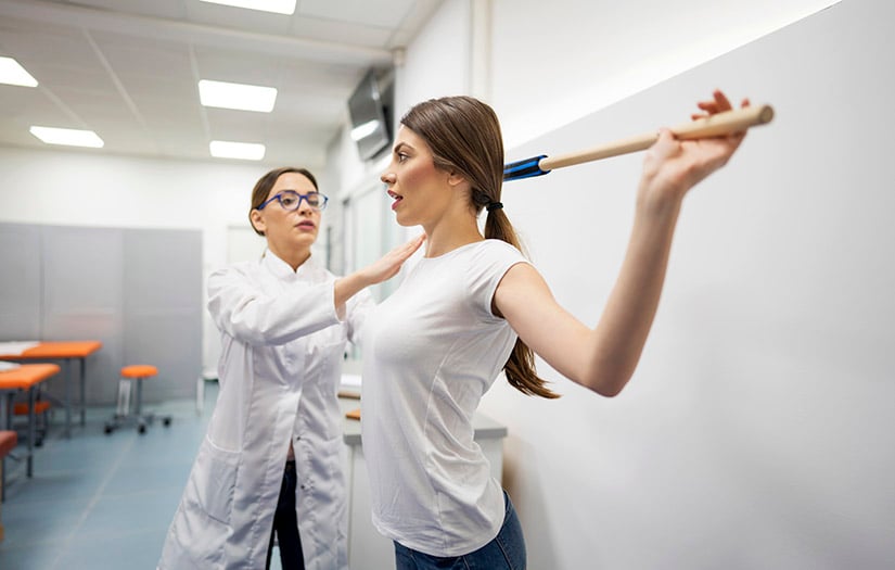
Postural and phasic muscle theory and its application takes a global view of body and joint movement. It’s about function, how the body is designed to perform and how to improve that function by working within the body’s design. If we understand function, we can problem solve and plan better programs for our clients’ individual needs. Learning and working with postural and phasic muscle applications is a good place to start.
As a therapist and teacher, I have used this concept for 14 years as a reliable assessment and rehabilitation tool. The techniques are field-oriented rather than research oriented and can be readily used in a fitness setting without the need for research equipment. Skills in hands-on assessment and observation are excellent tools for this kind of work.
The relationship of postural/phasic muscles offers a strategy for conditioning. Instead of randomly stretching and/or strengthening muscles, postural/phasic theory teaches the trainer a hierarchy of exercises that support the body in its “bipedal challenge” as well as program development for individual client goals. Good function and posture is frequently based on certain postural/phasic muscle relationships. Trainers can use this kind of information to develop programs and get better, faster results with clients.
In this article, I will present the taxonomy of postural/phasic muscles, their characteristics and reciprocal relationships. I will also discuss several applications and stretches.
For Corrective Exercise Specialists (and other fitness professionals alike) the strategies used within this article will help take your practice to the next level.
The Source of Postural/Phasic Theory
In the late 1960s, Dr. Vladimir Janda, a Czechoslovakian physiatrist and physiotherapist, determined variations in muscles using EMG and clinical studies. He named certain of these muscles “postural” because of their tonic or anti-gravity behavior. Other muscles he called “phasic.”
Phasic muscles are available on demand but are not responsive to the pull of gravity. He also found that muscles influence each other when they are opposed in the body, beyond agonist/antagonist pairings. The postural/phasic relationship is “a natural, physiologically balanced imbalance between two systems.” Thus, muscles are not created equal. Variations in structure, function and physiology are well known.
The chart below outlines the characteristics and “balanced imbalance” of postural and phasic muscles.
| POSTURAL MUSCLE CHARACTERISTICS | PHASIC MUSCLE CHARACTERISTICS |
| Are anti-gravity or tonic muscles; they have a higher resting tonus than phasic muscles | Are available on demand but do not oppose gravity |
| Tend toward shortness and tightness | Tend toward inhibition and weakness |
| Are genetically older and less reactive to injury | Are genetically younger and more reactive to injury |
| Atrophy less quickly than phasic muscles | Atrophy more quickly than postural muscles |
Postural Muscles Definition
Muscles in the postural group are inherently active to help us oppose gravity and keep us on our feet, particularly on one foot during the gait cycle. Being in constant use may contribute to postural muscles’ higher resting tonus, readiness to act and also to a tendency to have a shorter than normal resting length.
Such shortening occurs even in normal activities and at rest. While the tendency of a muscle to be short or tight is not fully understood, the pattern of such muscle behavior has been consistently observed.
Phasic Muscles Definition
Phasic muscles also generate a pattern in the body, one of weakness and inhibition. These muscles are not necessarily injured, but they are insufficiently active and may even exhibit a kind of false paralysis. Muscles in this category include the abdominals, the gluteals, the deep neck flexors and the rhomboids (see a complete list below). These muscles seldom act alone but typically act in relation to postural muscles.
Consider, for example, the postural pattern of a hyperkyphotic thorax with rounded shoulders, depressed chest and forward head. This “upper crossed syndrome” is a product of shortened pectoralis major fibers and cervical extensors paired with weak deep neck flexors, rhomboids, middle trapezius and serratus anterior, further complicated by a static immobility in the erector spinae. No one muscle is at fault. All of them are dysfunctional and should be addressed as a series of interacting forces.
Testing all of the postural muscles in the body (listed below) will generate a stretching plan for use within the fitness program. A single postural muscle/group such as the hamstrings can be tested and stretched as needed. While we can lengthen a postural muscle by isolated stretching, we cannot effectively strengthen a phasic muscle in isolation.
Doing so may weaken the muscle further and result in a poor movement pattern. Consider the current emphasis on “core” strength and stability. The phasic abdominal muscles tend toward weakness. Conventional training frequently tries to strengthen the abdominals in isolation, forgetting they are paired with shortened postural muscles (ie.., the erector spinae). I will return to this topic later.
It is understandable that muscles in humans function as postural and phasic when we think about what I have already mentioned as the “bipedal challenge.” We are the only creatures on earth who develop to stand and walk on two feet, who walk on the whole foot and must organize the bipedal stance around a center of gravity that is always in motion, even when we are “just” breathing.
Standing and walking with the body in good alignment is a nearly effortless process due to the give and take of muscular activity and a finely tuned proprioceptive system. However, innate functional patterns in the CNS are skewed by sedentary life, insufficient variety of body movement and/or random training on a poor foundation.
Postural VS. Phasic Muscles
| Muscles with mainly POSTURAL function | Muscles with mainly PHASIC function |
| Sternocleidomastoid | Longus colli and capitis |
| Upper fibers of trapezius | Scaleni (varies from postural to phasic) |
| Levator scapulae | Pectoralis major (abdominal fibers) |
| Flexors of the upper extremity: pectoralis major (clavicular and sternal fibers); anterior deltoid, long head of biceps | Extensors of the upper extremity: posterior deltoid, teres major, latissimus dorsi |
| Quadratus lumborum, erector spinae group, rotatores, multifidi | Middle and lower fibers of trapezius |
| Hip flexors: iliopsoas, rectus femoris, TFL | Subscapularis |
| Piriformis | Romboids |
| Hip adductors (one-joint muscles only) | Serratus anterior |
| Hip extensors: all three hamstring muscles | Abdominals: rectus , internal and external obliques |
| Plantar flexors: gastroc, soleus, tibialis posterior | Gluteus maximus, medius, minimus |
| Vastus medialis and lateralis | |
| Tibialis anterior | |
| Peronei (fibulari) |
The above chart does not list all of the muscles in the body. It lists only the muscles identified by Dr. Janda in the two categories of postural and phasic activity with respect to the gravitational stresses of a bipedal stance. The chart is organized vertically from the head of the body to the feet. There is no intended relationship among muscles that appear on the same horizontal line.
The listing is a compact guideline useful for learning the postural and phasic muscles in the body and for organizing an assessment. It forms the basis for a systematic stretching protocol. The trainer benefits with an orderly, manageable and finite number of muscles to check and to track for change.
Postural muscle testing and stretching reminds us that stretching is more than a momentary recuperation from resistance work. It is an essential process for maintenance of specific muscle groups and body-level relationships. Stretching is a skill that deserves to be well taught and when learned, becomes a life-long tool for well being. Stretching has many benefits, notably reducing the discomfort of limited ROM and enabling improved performance in sport, work and daily activity.
Case Study: Gait Limitations in a Postural/Phasic Context
A female client used to enjoy walking but now finds it exhausting and too much work for too little result. She reports low back and/or sacroiliac pain. Observing her walk, we see a shuffling gait, a hip joint that does not easily extend and a poor propulsion pattern (push off with the toes). Tests of her postural hip flexors reveal a shortened iliopsoas, rectus femoris and tensor fasciae latae. The gastroc/soleus group is also shortened.
Based on postural/phasic protocol, we stretch the client’s tight hip flexors and gastroc/soleus and provide an interim program of self stretches. We avoid strengthening at this stage because weakness is not her main problem.
Over a period of two or three sessions, her gait pattern improves and her pain is reduced or eliminated. The stretches got her back on her feet and able to enjoy walking again. She returns for a new phase of conditioning.
Analysis
We stretch the postural muscles first because they are inhibiting hip extension and restricting plantar flexion. Hip flexors must eccentrically contract to allow hip extension, but if shortened, they will not be able to lengthen adequately and will inhibit extension and propulsion in the gait pattern. In this example, pain is a product of what Janda called a “trick” movement or a substitute of other than prime movers to achieve the desired goal.
The poor gait pattern is also a kind of “trick.” The client is asked to walk, and lacking good neuromuscular resources, she uses whatever muscles are available. Her shuffling gait and limited stride cannot be voluntarily corrected without introducing more tricks. The CNS (which is intention-driven) tries to get hip extension from the lumbar spine or from rotation in the pelvis. Such inappropriate use of the body often produces pain in the stressed areas. This pain is secondary to the dysfunctional extension of the hip joint.
Effective Stretching
To work with postural/phasic theory, it is essential to have good stretching techniques and teaching skills and to visualize the functional pathway of a muscle. In the above example, the rectus femoris is the only postural muscle of the quadriceps group. As a biarticular muscle, the stretch for it must take into account both the hip and the knee joints.
(The familiar heel-to-buttock stretch applies only to the short quads.) The long quads require a kneeling (Figure 1) or side-lying position (Figure 2) in which, in its ideal configuration, the hip joint is at least in neutral extension and the knee joint flexed to 90 degrees. The stretch should be held for at least 30 seconds, performed at least twice and be supervised in the learning stage. Trainers should coach their clients to gradually develop the full resting length in this intensive stretching exercise.
The Taxonomy of Phasic Muscles
What else can we learn from this taxonomy of muscles? A deeper understanding of weakness and strength. The inability to move is often attributed to weakness when it may instead be a problem of chronic shortening. A muscle with a shortened resting length has neither optimal concentric or eccentric action. Because it lacks the give and take of normal muscle tissue, its ability to exert force is restricted.
The long back muscles (i.e., erector spinae, postural) are an excellent example. If they are tight, back extension will be limited but so will back flexion. This is an example of a limitation in movement caused by shortened rather than weak muscles. Janda describes this phenomenon as “tightness weakness.” The shortened postural muscle’s inability to deliver an adequate contraction exhibits weakness due to tightness. If the trainer tests only for strength and not length, the training protocol will be less effective.
When to Stretch, When to Strengthen
There is a hierarchy or at the very least an optimal protocol regarding the order in which we stretch and strengthen the muscles. Janda suggests the following plan: length, strength and endurance.
First, achieve an optimal resting length based on standard testing protocols, assisted stretching and self stretching.
Secondly, strengthen the muscle/groups, if necessary watching for any “trick” movement patterns.
Thirdly, increase endurance, muscle mass and definition by adding load, repetitions and sets based on individual goals. Finally, maintain normal resting length with a maintenance program of stretching.
Follow the chart listing the postural and phasic groups. Never try to strengthen postural muscles without first testing for shortness. Most of them are already relatively strong or may exhibit “tightness weakness.” Never try to strengthen phasic muscles without normalizing their opposing postural muscles (see the chart below for a examples of these reciprocal relationships).
While any muscle may become functionally tight, a phasic muscle will not be routinely tight. A postural muscle, on the other hand, is likely to be short and require stretching. This information tells us that we do not have to stretch every muscle in the body, but we should always test the postural muscles. Don’t stretch randomly and don’t generalize from one person to the next. There are always surprises.
Postural/phasic theory also teaches us about relationships in the body. The dominant pull of a postural muscle against an opposite phasic muscle may show itself as a postural deviation or a movement dysfunction. The chart below shows an array of such opposing muscle groups:
| POSTURAL MUSCLES | PHASIC MUSCLES |
| Posterior neck muscles (upper trapezius, levator scapulae, deep cervical extensors) | deep anterior cervical flexors (longus colli and capitis) |
| Quadratus lumborum, erector spinae, multifidi and rotatores | abdominal obliques and rectus |
| Rectus femoris, tensor fasciae latae, iliopsoas | gluteus maximus, medius, minimus |
| Pectoralis major (sternal and clavicular fibers) | rhomboids, middle trapezius, serratus anterior |
Phasic Muscles and Core Strength
Let’s look at an example of one such relationship by returning to the topic of “core” strength. Suppose you have a client who, in spite of regular abdominal strengthening exercises, fails to be able to do a complete, smooth and controlled abdominal curl. Look at the postural/phasic muscle relationships. If the postural erector spinae are tight, the phasic abdominals, flexors of the trunk, will not be able to override the back extensors in the exercise.
The client’s abdominal curl will be incomplete or forced and will not change despite diligent practice. The client may give up in frustration. Furthermore, because the abdominals are ineffective for the task, the CNS will attempt to substitute other muscles such as the superficial neck flexors, iliopsoas or the back extensors (lumbar hyperextension) to do the work.
By first stretching the back muscles, the abdominals will be able act on demand, and your client will have a chance to develop a strong abdominal wall. Janda repeatedly demonstrated this in his research. I have used this principle in my rehabilitation practice and teaching for many years and have found it consistently applicable.
At the June 2002 Canadian Personal Trainers Network conference in Toronto, I taught my 3-D Back Stretch to a group of 85 trainers. Prior to learning the stretch, each trainer did 10 abdominal curls as a pre test. Following the stretch, each trainer repeated the curls as a post test. Almost all reported changes such as increased symmetry of movement, smoother action, longer retention of abdominal flexion and ease of descent.
References:
Janda, V (1994). Muscles and motor control in cervicogenic disorders. In Grant, R (ed). Physical therapy of the cervical and thoracic spine. Edinburgh: Churchill Livingstone.
Janda, V (1986). Muscle weakness and inhibition (pseudoparesis) in back pain syndromes. In Grieve, GP (ed). Modern manual therapy of the vertebral column. First ed. Edinburgh: Churchill Livingstone.
Janda, V (1983). Muscle function testing. London: Butterworths.
Janda, V (1978). Muscles, central nervous motor regulation, and back problems.
In Korr, IM (ed). The neurobiologic mechanisms of manipulative therapy. NY: Plenum Press.
Jull, G and V Janda (1987). Muscles and motor control in low back pain. In Twomey, LT and Taylor, JR (eds). Physical therapy of the low back. 1st ed. NY: Churchill Livingstone.
Tunnell, PW (1998). Muscle length assessment of tightness prone muscles. Journal of Bodywork and Movement Therapies, 2:1, 21-26.
Woodruff, DL (2002). Postural muscle assessment and stretching techniques. Course manual. By the author.
Woodruff, DL (2001). 3-D workout, vol. 1: the basics. VHS videotape, 48 minutes. Oakville, ON: Body-in-Motion.


